|
|
|
| |
| ABSTRACT |
|
The aim of this study was to analyze the relationship of range of motion (ROM) in the sagittal plane and timing parameters during a bodyweight squat to the depth of the squat. Sixty participants (20 females and 40 males) took part in this study. They were instructed to perform a bodyweight squat to the maximal depth position. Kinematic data were obtained using the optical motion capture system. The time for the descent phase of squatting was normalized from 0% (initial position, start of movement) to 100% (squat position-stop of movement). The ROM of ankle, knee, hip, pelvis and spine in the sagittal plane and the normalized time when the maximum joint angles occurred during the descent were analyzed to investigate the relationship between them and the squat depth in males and females. The knee ROM contributed most significantly, from all joints to squatting depth in both females and males (r = 0.92, p < 0.001). The squat depth was related to lumbar, hip and knee motion in females and to all kinematics parameters in males. Maximal ankle dorsiflexion and pelvis anterior tilt were reached earlier than the maximal angles of knee, hip and spine during squatting. Pelvis and ankle timing was negatively correlated with the squat depth (rs = -0.64, p < 0.001 and rs = -0.29, p = 0.02, respectively). This suggests that pelvis and ankle timing can be important to keeping balance during squatting and can lead to achieving the desired depth. |
| Key words:
Squat depth, lumbar spine, pelvis, ankle, kinematics
|
Key
Points
- This study aimed to analyze the relationship of range of motion (ROM) in the sagittal plane and timing parameters to the squat depth during a bodyweight squat.
- Pelvis and ankle timing was negatively correlated with the squat depth, which suggests that pelvis and ankle timing can be important in keeping balance during squatting and can lead to achieving the desired depth of the squat.
- During squatting maximal ankle dorsiflexion and pelvis anterior tilt were reached earlier than the maximal angles of knee, hip and spine.
|
Squatting is widely used in physical therapy and sport training as a closed-kinematic chain exercise involving all lower limb joints and many muscles. It is one of the most important elements in building leg strength. Due to its multi-joint character, squatting seems to be an evaluation tool in motor control assessment (Gawda et al., 2019). Factors affecting squatting techniques are considered crucial in avoiding overuse or injury. They have aroused great interest among researchers in recent years (Lee et al., 2015; Zawadka et al., 2018). One of the variables influencing muscular effort during squatting is depth (Bryanton et al., 2012). A full-range-of -motion (ROM) squat and a squat with limited depth are both common exercises of professional and recreational athletes. They demand different power and force and cannot be used interchangeably in weight training (Drinkwater et al., 2012; Kitamura et al., 2019; Martínez-Cava et al., 2019). A squat requires mobility of the lower limb joints and the trunk. Although movement is always three dimensional, squatting involves mainly motion in the sagittal plane. Thus, squat depth can be defined using the knee flexion angle (Bryanton et al., 2012) or the descent expressed as percentage of leg length (Bagwell et al., 2016; Kim et al., 2015). However, no standardized measures have been universally recognized, and terminology can differ among researchers (Schoenfeld, 2010). Previous studies investigated the passive and active ROM of lower limbs and correlated it with the depth or the kinematics of the squat (Drinkwater et al., 2012; Kim et al., 2015). It was reported that a deep squat can be a screening test to assess bilateral symmetrical mobility of the hip, knee, and ankles (Cook et al., 2014; Kritz et al., 2009). Previous studies have suggested that the weakness or poor mobility of joints are factors limiting successful squatting performance (Escamilla, 2001; Macrum et al., 2012; Zawadka et al., 2018). Squat as a multi-joint, closed kinematic chain exercise requires the coordinated movement of many segments (Fuglsang et al., 2017; Howe et al., 2019). Their mobility can affect adjacent joints motion during exercise. Thus, there is still a need to investigate the relationship between a squatting movement pattern and its depth to establish the normative parameters of a motion. Moreover, research also suggests that sex differences should be considered when developing trunk geometry (Marras et al., 2001) and when analyzing kinematics and timing during exercise and functional tasks (Graci et al., 2012; Thomas et al., 1998). Alterations in movement patterns during functional tasks are considered as one of the factors contributing to a greater risk of musculoskeletal disorders and lower limb injuries (Powers, 2003, 2010). Understanding a squat movement pattern and its range can be important for coaches and physical therapists during the assessment and teaching of bodyweight squat techniques. Nevertheless, the effect of sex and different squat depths on the lower limb and spine kinematics still remain unclear in many aspects. One of them is the relation of a joint’s movement timing to squat performance. McKean et al. previously investigated the movement pattern of back squats in males and females showing significant differences in lumbar- sacral coordination and ranges but they did not measure squat depth (McKean et al., 2010b). In spite of the fact that coordination and timing aspects of motion have been well explored, to the best of the authors’ knowledge, there have not been any previous reports investigating the relation between timing parameters and squat depth. Therefore, the purpose of this study is to investigate the relationship between squat depth and range of motion in the sagittal plane and timing parameters during a bodyweight squat in both males and females. We hypothesize, based on the findings of previous studies, that all joints will achieve maximal angles almost simultaneously with the deepest part of the squat and that the knee ROM will be the most strongly related to squat depth from all kinematic variables. However, we expect to observe differences between males and females in squat movement patterns. SubjectsSixty healthy, university-age, recreationally active volunteers took part in this study. The participants were not especially trained in squatting. Subjects were excluded if they reported neurological signs, lower extremity or back pain, a history of lower extremity or back injury or surgery. In all cases participants gave their written informed consent. Table 1 shows characteristics of the groups. Ethics approvals were obtained from the local university Bio Ethics Committee (approval number KE-0254/322/2018).
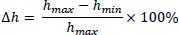
ProceduresRecreationally active, healthy volunteers were instructed to squat without additional load to their maximum depth. Each of them performed a full range of squat motion using their usual technique. Kinematic data (ROM’s of spine, pelvis, hip, knee and ankle in the sagittal plane) and timing of when maximum angles of each joint were achieved during squat descent were collected and analyzed. Squat depth was defined as a leg length using the height of a marker placed on the second sacral spinous process. The participants were instructed to perform a bodyweight squat to the maximal depth position they were able to hold for 3s. The depth was controlled and had to be equal or greater than 30% of leg length without causing any pain. The initial position of participants was specified as upright standing, looking straight ahead with feet at shoulder width apart and arms extended forward and parallel to the floor. The participants were asked to descend, hold their position and then, to ascend and return to their initial position. They were instructed to maintain heel contact throughout the task. During the first step, no more than 3 squat trials were made to practice and avoid fatigue. Afterwards, 5 repetitions were performed and kinematic data were collected. Finally, 3 middle trials (without the first and the last) were averaged and further analyzed. Because the aim was to analyze natural movement patterns, the squat technique was not imposed except with the guidelines mentioned above. Therefore, any mistakes in technique and signs of compensation were not corrected. Participants performed the exercise at their preferred pace. Leg length was defined as the height of a marker placed on the second sacral spinous process (Figure 1.). Further, the squat depth was calculated according to the following equation:
Δh-descent
hmax-the highest position of S2 marker
hmin-the lowest position of S2 marker
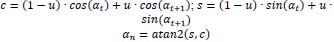
Kinematic data were obtained using the VICON motion analysis system (Oxford Metrics Ltd, Oxford, UK) with eight near-infrared cameras operating at a sampling rate of 100 Hz. Joint angles were defined as: the angle of the shank relative to the foot (ankle angle), the angle of the thigh relative to the shank (knee angle), and the angle of the pelvis relative to the thigh (hip angle), the angle of the pelvis relative to the global coordinate system (pelvis angle) and the angle of the thoracic spine relative to the pelvis (lumbar spine angle). An XYZ Cardan sequence was used. Markers placed according to the Plugin-gait model and additional markers placed for pelvis and lumbar spine angles calculation are presented in Figure 2. Motion data were first processed and analyzed in Nexus software (version 2.9.1) using the standard 3D-rigid-body-linked–segment-modelling procedures and modelling procedures based on additional markers. Data processing was partially automated using a custom c3d file processing tool. The tool utilizes Eigen and Biomechanical Toolkit libraries. The automatic data processing flow consisted of the following steps: (1) trial information, including the beginning and ending frames of descent and ascent squat phases, was read from a file, (2) data recorded in all trials were verified (presence of required markers was checked in the desired time ranges), (3) angle data in each phase were normalized and expressed as a percentage of phase duration, (4) minimal and maximal values of angles of interest, squat depth, normalized times of their occurrence were found, (5) results were saved to a file. Resampling consisted of computing interpolated values for the requested normalized time moments using their neighboring values from c3d files. For interpolating angular values the following formulas were used:
Where: αt and αt+1 – original angle values for times t and t+1, u - division ratio of [t,t+1] time range by the normalized time moment n, atan2 – a popular variant of the arctangent function implementation.
The time for the descent phase was normalized from 0% (initial position, start of movement) to 100% (squat position-stop of movement). The results reported in this study show the timing of when maximum angles of each joint were achieved during the descent phase of motion. Maximum angles mean maximal: pelvis anterior tilt, flexion of hip, flexion of knee and flexion of lumbar spine and ankle dorsiflexion. ROM was defined as the difference between the maximal and minimal angle obtained in the sagittal plane during squatting. In addition, the contribution of each joint ROM in the total ROM of the kinematic chain was calculated. Total ROM was defined as the sum of all joints’ ROMs during squatting.
Statistical analysisData analysis was conducted using the Statistica software (ver. 13.1). The significance level was set at p = 0.05. For normally distributed data the independent t-test was used. For non-normally distributed data the U-Mann Whitney test was used to determine differences in timing between males and females. Effect sizes were determined using the Cohen’s d coefficient and the Glass rank-biserial correlation coefficient. Cohen’s coefficient was interpreted as: small (0.2-0.5), moderate (0.5-0.8), or large (>0.8) and Glass coefficient as: small (0.1-0.3) moderate (0.3-0.5.) and large (>0.5). The Pearson correlation was used to examine the relationships between the ROM of the lumbar spine, pelvis, hip, knee and ankle achieved in the squatting position and the squat depth. The Spearman rank correlation was used to examine the relationships between the timing parameters and the squat depth. The analysis was carried out for all participants collectively and separately, for men and women. The intra-class correlation coefficients among the three repetitions show excellent reliability: ankle - 0.94; knee - 0.92; hip - 0.90; pelvis - 0.90; lumbar spine - 0.93. Graphs were prepared using Microsoft Office Excel.
A comparison of males and females revealed that only the lumbar spine ROM showed a significant difference between the groups. Males demonstrated a greater lumbar spine ROM than females during their maximal squatting (43.74 ± 15.31 deg vs 30.08 ± 14.44 deg respectively; p < 0.01). Other kinematic variabilities were not statistically different when comparing males and females However, analysis of each joint contribution in the total ROM in the sagittal plane demonstrated that females had a greater contribution of pelvis ROM during squatting and a smaller contribution of lumbar spine ROM than males (pelvis: 7.47 % ± 2.50 vs. 5.94% ± 2.59, p = 0.03; lumbar spine: 9.24% ± 3.99 vs. 12.89 % ± 3.97, p = 0.001). The U-Mann Whitney test revealed a difference in normalized time when the maximum angle of the pelvis and spine occurred among males and females (p = 0.02) with moderate effect size. The maximal pelvis angle was achieved earlier by males than by females (at 62.33% and 70.25% respectively), and the maximal spine angle was obtained by males slightly later than by females (at 98.83% and 97.58% respectively). Other variabilities were not statistically different when comparing males and females (Table 2 and Table 3). There were moderate to very high (r = 0.49-0.92) positive correlations between ROMs of the lumbar spine, hips, knee and squat depth in the group of females. The pelvis and ankle ROMs were not related to squat depth in females (r = 0.14, p = 0.56; r = 0.43, p = 0.06, respectively). In males, the ROMs of hip, pelvis, ankle, lumbar spine and knee were weakly to very strongly (r = 0.32-0.92) correlated with the maximal squat depth. With no division into groups, the maximal squatting depth was related with the knee ROM (r = 0.96; p < 0.001), the lumbar spine ROM (r = 0.61; p < 0.001), the hip ROM (r = 0.45; p < 0.001) and the ankle ROM (r = 0.38, p = 0.003) (Table 4, Figure 3). There was negative correlation between pelvis timing and squat depth in both females (r = -0.48; p = 0.03) and males (r = -0.71; p < 0.001). There was also a weak negative correlation in males between the ankle timing and squat depth (r = -0.33; p = 0.04) (Table 5). Range of motionThe purpose of this study was to analyze how the ROMs of the lower limbs’ joints, pelvis and lumbar spine were related to squat depth when the squat was performed by untrained participants (men and women). Males and females demonstrated differences in kinematics and timing during squatting. It is worth noticing that, despite the similar average squat depth in both males and females and similar average ranges observed in the joints of lower limbs and pelvis, males showed significantly greater lumbar spine ROM than females did. Moreover, females had a greater contribution of pelvis ROM during squatting and a smaller contribution of lumbar spine ROM than males The movement pattern used by females when squatting involves a much stiffer lumbar spine position. A more flexed posture during squatting can generate greater stress to passive tissues of the spine in males. This finding is consistent with the findings of McKean et al (McKean et al., 2010b). However, in the McKean study the depth of the squat was not controlled. Moreover, only the sacrum and lumbar spine motion was investigated by McKean et al. The sex-dependent difference in kinematics during squatting should be considered in training programs of recreational athletes. Another finding of the present investigation is that the knee ROM contributed most significantly to squatting depth from all joints in both males and females. As expected, the squat depth seems to be determined mostly by knee kinematics. Thus, the knee angle of flexion during squatting is recommended as a parameter for describing squat depth very well. Other valuable results from this study are: (1) squat depth was not correlated to pelvis and ankle ROMs in females and (2) squat depth was correlated to all investigated ROMs in males. It had been previously reported that there are significant differences in movement patterns between female and males during exercise performance (McKean et al., 2010a, 2010b). Ankle mobility was considered in many studies to be crucial to keeping a proper movement pattern during squatting and squat depth (Dill et al., 2014; Macrum et al., 2012). Kim et al. reported that the ankle dorsiflexion ROM with flexed and extended knee measured by a goniometer correlated significantly with squat depth in male and female subjects (Kim et al., 2015). However, analysis of the strongest correlations suggest that squat depth in females depends mostly on knee and hip kinematics and in males depends on knee and spine kinematics. These dissimilarities between the sexes should be considered during deep squats training.
TimingMales reached their maximal angle of pelvis significantly earlier than females in this study. The spine maximal angle was reached by males slightly later than by females. Nevertheless, the order in which maximal angles are reached is similar for both genders. First, the pelvis reaches its maximal anterior tilt and the tilt stays constant or decreases for the rest of the descent phase. Second, the ankle reaches its maximal angle of dorsiflexion and after that the spine, the knee and the hip reach their maximal angles almost simultaneously at the end of the descent phase. This is consistent with the findings of McKean et al., who showed a similar relation in the timing of lumbar flexion (McKean et al., 2010b) This author also reported that maximum hip and knee angles are achieved almost simultaneously with the deepest part of the squat (M. McKean and Burkett, 2012). To the best of the authors’ knowledge, this is the first report of a correlation between the timing parameters of joints’ motion and squat depth. We have shown that increased squat depth is related to the pelvis reaching the maximal angle of anterior tilt earlier during the squat. It is probably caused by the retroversion of the pelvis in a deeper squat position. Also, the ankle dorsiflexion in males was reached earlier when the depth of squat increased. Other joints show a similar coordination without relation to squat depth. These two joints (pelvis and ankle) clearly show a different ability to adjust to increasing squat depth as compared to other joints. It is well known to coaches and physical therapists that the ankle and pelvis motion restriction can be a cause of compensatory motion in adjacent segments (Dill et al., 2014; Macrum et al., 2012). For this reason, both kinematic and time-related parameters variables of these elements should be considered during examination for sports training and medical diagnosis. A possible explanation of our results could be the controlling role of the pelvis and ankle in squatting performance (Fuglsang et al., 2017). Once the pelvis/ankle reaches its maximal angle, the angle remains constant (the plateau is maintained) or the motion can occur in the opposite direction. The mechanism present in a specific moment of movement can be important in keeping balance during squatting and can lead to achieving the desired depth. Interestingly, retroversion of the pelvis at the deepest point of the squat seems to be important in characterizing a good execution of squatting (Bagwell et al., 2016; Lamontagne et al., 2009) However, these hypotheses require further research. Moreover, pelvis and ankle demonstrate a smaller ROM than other segments in the sagittal plane during squatting and therefore have limited ability to adjust to increasing squat depth.
Practical ApplicationsThis study may provide additional knowledge for improving teaching and monitoring of squat techniques. Moreover, results of this study can establish guidelines for movement coordination in the sagittal plane of the lower limbs and the lumbar spine. Such guidelines could be used as a standard for squatting. Examination of movement patterns, as our study shows, should be done very carefully due to significant differences that exist in the manner in which men and women perform the squat. Females showed a greater contribution of pelvis ROM and a smaller contribution of lumbar spine ROM in squatting than males. Because this finding is consistent with previous reports, it may be considered as a typical way of squat executing by young males and females. The ROM of each joint in the sagittal plane generally, except the pelvis in females, seems to increase with the squat depth. On the other hand, pelvis timing showed to be significantly correlated with squat depth in both males and females, ankle timing was correlated with squat depth in males. Coaches and therapists should consider that knees, hips and lumbar spine reached their maximal angles almost at the deepest point of the squat no matter how deep the squat was. However, they should also consider that deeper squat is related to the earlier achievement of pelvis and ankle maximal angles. Exercises that increase joints mobility may be beneficial in improving squatting, but right timing should also be included in training practice. We recommend greater attention to the pelvis-ankle regulatory mechanism in keeping a balanced movement pattern of the deep squat.
LimitationsFirst, we are unable to generalize our findings to other age groups. All subjects participating in this study were in their early twenties. A future study might also examine experienced weight lifters. Second, we focused only on the ROM in the sagittal plane. A compensatory mechanism can also occur in the frontal and transverse planes, especially in the knee and ankle (Lee et al., 2015; Macrum et al., 2012). However, we consider the sagittal plane motion as the most important factor during a squatting descent. Third, the ROM in joints was not tested in this study in the way it is normally executed during a physical examination using a goniometer. However, the ranges observed in this study can be considered as functional ROMs for joints during squatting (Rabin and Kozol, 2017). We are also aware that sex-dependent differences in the kinematics might also be due to the anthropometric differences since males displayed a greater body weight and body height than females. Results of this study should be interpreted bearing in mind that the sample size was small (which is a common problem in motion capture investigations) and the numbers of males and females were not equal.
The current study has demonstrated that the knee ROM contributed most significantly of all joints to squatting depth in both males and females. Movement patterns are different for females and males during the requested squat. Differences in kinematics and timing between the sexes should be considered by coaches and therapists when they instruct untrained persons about bodyweight squatting. During squatting maximal ankle dorsiflexion and pelvis anterior tilt are reached earlier than maximal angles of the knee, hip and lumbar spine. Pelvis and ankle kinematics are generally weakly affected by squat depth, but their timing is related to this parameter. When increasing the squatting depth, the normalized time when the pelvis and the ankle reach maximal angles is earlier during the squat. This suggests that pelvis and ankle timing can be important in keeping balance during squatting and can lead to achieving the desired depth of the squat.
| ACKNOWLEDGEMENTS |
This research did not receive any specific grant from funding agencies in the public, commercial, or not-for-profit sectors. The author declare that they have no conflict of interest. The experiments comply with the current laws of Poland. |
|
| AUTHOR BIOGRAPHY |
|
 |
Magdalena Zawadka |
| Employment: Department of Sports Medicine, Faculty of Health Sciences, Medical University of Lublin, Chodzki 7 Street, 20-093 Lublin, Poland |
| Degree: MSc |
| Research interests: Biomechanics, physical therapy in sport |
| E-mail: magdlenzawadka91@gmail.com |
| |
 |
Jakub Smolka |
| Employment: Department of Computer Science, Faculty of Electrical Engineering and Computer Science, Lublin University of Technology, 20-618 Lublin, Nadbystrzycka 38D Poland |
| Degree: PhD |
| Research interests: motion capture, image processing, applications of mobile devices |
| E-mail: jakub.smolka@pollub.pl |
| |
 |
Maria Skublewska-Paszkowska |
| Employment: Department of Computer Science, Faculty of Electrical Engineering and Computer Science, Lublin University of Technology, 20-618 Lublin, Nadbystrzycka 38D Poland |
| Degree: PhD |
| Research interests: Motion analysis, biomechanics |
| E-mail: maria.paszkowska@pollub.pl |
| |
 |
Edyta Lukasik |
| Employment: Department of Computer Science, Faculty of Electrical Engineering and Computer Science, Lublin University of Technology, 20-618 Lublin, Nadbystrzycka 38D Poland |
| Degree: PhD |
| Research interests: Motion analysis, biomechanics |
| E-mail: e.lukasik@pollub.pl |
| |
 |
Piotr Gawda |
| Employment: Department of Sports Medicine, Faculty of Health Sciences, Medical University of Lublin, Chodzki 7 Street, 20-093 Lublin, Poland |
| Degree: PhD |
| Research interests: Sports medicine |
| E-mail: piotr.gawda@umlub.pl |
| |
|
| |
| REFERENCES |
 Bagwell J. J., Snibbe J., Gerhardt M., Powers C. M. (2016) Hip kinematics and kinetics in persons with and without cam femoroacetabular impingement during a deep squat task. Clinical Biomechanics 31, 87-92. |
Bryanton M. A., Kennedy M. D., Carey J. P., Chiu L. Z. F. (2012) Effect of Squat Depth and Barbell Load on Relative Muscular Effort in Squatting. Journal of Strength and Conditioning Research 26, 2820-2828. |
Cook G., Burton L., Hoogenboom B. J., Voight M. (2014) Functional movement screening: the use of fundamental movements as an assessment of function - part 1. International Journal of Sports Physical Therapy 9, 396-409. |
Dill K. E., Begalle R. L., Frank B. S., Zinder S. M., Padua D. A. (2014) Altered knee and ankle kinematics during squatting in those with limited weight-bearing-lunge ankle-dorsiflexion range of motion. Journal of Athletic Training 49, 723-732. |
Drinkwater E. J., Moore N. R., Bird S. P. (2012) Effects of changing from full range of motion to partial range of motion on squat kinetics. Journal of Strength and Conditioning Research 26, 890-896. |
Escamilla R. F. (2001) Knee biomechanics of the dynamic squat exercise. Medicine and Science in Sports and Exercise 33, 127-141. |
Fuglsang E. I., Telling A. S., Sørensen H. (2017) Effect of Ankle Mobility and Segment Ratios on Trunk Lean in the Barbell Back Squat. Journal of Strength and Conditioning Research 31, 3024-3033. |
Gawda P., Ginszt M., Zawadka M., Skublewska-Paszkowska M., Smołka J., Łukasik E., Majcher P. (2019) Bioelectrical Activity of Vastus Medialis and Rectus Femoris Muscles in Recreational Runners with Anterior Knee Pain. Journal of Human Kinetics 66, 81-88. |
Graci V., Van Dillen L. R., Salsich G. B. (2012) Gender Differences in Trunk, Pelvis and Lower Limb Kinematics During a Single Leg Squat. Gait & Posture 36, 461-466. |
Hartmann H., Wirth K., Klusemann M. (2013) Analysis of the Load on the Knee Joint and Vertebral Column with Changes in Squatting Depth and Weight Load. Sports Medicine 43, 993-1008. |
Howe L. P., Bampouras T. M., North J., Waldron M. (2019) Ankle dorsiflexion range of motion is associated with kinematic but not kinetic variables related to bilateral drop-landing performance at various drop heights. Human Movement Science 64, 320-328. |
Kim S.-H., Kwon O.-Y., Park K.-N., Jeon I.-C., Weon J.-H. (2015) Lower Extremity Strength and the Range of Motion in Relation to Squat Depth. Journal of Human Kinetics 45, 59-69. |
Kitamura T., Kido A., Ishida Y., Kobayashi Y., Tsukamoto S., Tanaka Y. (2019) Muscle Activity Pattern with A Shifted Center of Pressure during the Squat Exercise. Journal of Sports Science and Medicine 18, 248-252. |
Kritz M., Cronin J., Hume P. (2009) The Bodyweight Squat: A Movement Screen for the Squat Pattern. Strength & Conditioning Journal 31, 76-85. |
Lamontagne M., Kennedy M. J., Beaulé P. E. (2009) The Effect of Cam FAI on Hip and Pelvic Motion during Maximum Squat. Clinical Orthopaedics and Related Research 467, 645-650. |
Lee J. D., Koh D. H., Kim K. (2015) The kinematics of the lower leg in the sagittal plane during downward squatting in persons with pronated feet. Journal of Physical Therapy Science 27, 285-287. |
Macrum E., Bell D. R., Boling M., Lewek M., Padua D. (2012) Effect of Limiting Ankle-Dorsiflexion Range of Motion on Lower Extremity Kinematics and Muscle-Activation Patterns during a Squat. Journal of Sport Rehabilitation 21, 144-150. |
Marras W. S., Jorgensen M. J., Granata K. P., Wiand B. (2001) Female and male trunk geometry: size and prediction of the spine loading trunk muscles derived from MRI. Clinical Biomechanics 16, 38-46. |
Martínez-Cava A., Morán-Navarro R., Sánchez-Medina L., González-Badillo J. J., Pallarés J. G. (2019) Velocity- and power-load relationships in the half, parallel and full back squat. Journal of Sports Sciences 37, 1088-1096. |
McKean M. R., Dunn P. K., Burkett B. J. (2010a) Quantifying the movement and the influence of load in the back squat exercise. Journal of Strength and Conditioning Research 24, 1671-1679. |
McKean M. R., Dunn P. K., Burkett B. J. (2010b) The lumbar and sacrum movement pattern during the back squat exercise. Journal of Strength and Conditioning Research 24, 2731-2741. |
McKean M., Burkett B. J. (2012) Does segment length influence the hip, knee and ankle coordination during the squat movement. Journal of Fitness Research 1, 23-30. |
Powers C. M. (2003) The Influence of Altered Lower-Extremity Kinematics on Patellofemoral Joint Dysfunction: A Theoretical Perspective. Journal of Orthopaedic & Sports Physical Therapy 33, 639-646. |
Powers C. M. (2010) The influence of abnormal hip mechanics on knee injury: a biomechanical perspective. The Journal of Orthopaedic and Sports Physical Therapy 40, 42-51. |
Rabin A., Kozol Z. (2017) Utility of the Overhead Squat and Forward Arm Squat in Screening for Limited Ankle Dorsiflexion. Journal of Strength and Conditioning Research 31, 1251-1258. |
Schoenfeld B. J. (2010) Squatting kinematics and kinetics and their application to exercise performance. Journal of Strength and Conditioning Research / National Strength & Conditioning Association 24, 3497-3506. |
Thomas J. S., Corcos D. M., Hasan Z. (1998) The influence of gender on spine, hip, knee, and ankle motions during a reaching task. Journal of Motor Behavior 30, 98-103. |
Zawadka M., Lukasik E., Skublewska-Paszkowska M., Smolka J., Gawda P., Jablonski M. (2018) Analysis of the kinematic parameters of squatting in subjects with different levels of physical activity - A preliminary study. Baltic Journal of Health and Physical Activity. The Journal of Gdansk University of Physical Education and Sport 10, 97-105. |
|
| |
|
|
|
|